Utilizing Geospatial Tools for Medical Image Analysis
Anonym
Remote Sensing has been around since the 1960s so not suprisingly, the science at its core has been altered and refined over time. Today it is fairly easy to utilize an image analysis program to see what shows up in an image taken today that wasn't in an image taken previously. If you want to know spectrally what is different from image A to image B, this can be done with a couple of clicks. Compare that to the early days where someone would need to manually compare images and hope their eyes were good enough to catch any visible changes. The same change is happening in the medical field where doctors and medical researchers are looking at ways to automate manual review processes with imagery. This is where geospatial analytic tools come into play.
Rather than reinventing the wheel and spending countless hours trying to write algorithms that mimic what is already being done with satellite imagery, doctors and medical researchers are looking to existing capabilities in the geospatial field to see how these tools can be repurposed. Over the past few years we have seen some very unique uses for ENVI, our image analysis software, in the medical field. One unique use as of late is researching whether ENVI and its change detection algorithms can used to detect early signs of skin cancer. The theory behind the idea was that with some manipulation to the images, they can be mosaicked to display a specific region of the body and then cataloged so when the patient visits next, the comparison can be run to see where new suspicious moles have cropped up or where changes in skin pigment have occurred. Those changes can be flagged for the doctor to review. With IDL’s DICOM support, it allows doctors to implement ENVI into a medical setting and push their patient data back up to servers where historical data is hosted, just as they would with traditional medical diagnostic tools. While there would be some challenges to getting complete body scans, the data itself could lead to finding trends on the skin that change how skin cancer is battled.
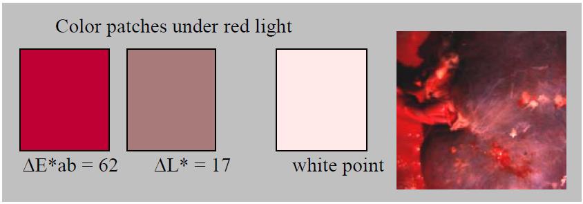
Another unique example of utilizing the existing geospatial capabilities from within ENVI for medical image analysis was done by National Institute of Standards and Technology. Their abstract focused on using a hyperspectral camera to study illuminant spectral distribution in surgical and diagnostics settings as well as how that affected human visual perception while performing surgery and diagnostics both in the body, as well as on the epidermis. The study took a look at both traditional and LED surgical lamp reflectance, compared to a spectrally tunable light source and what benefits the latter could potentially provide in both diagnostic and surgery settings.
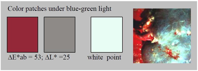
Illumination with enhanced contrast as a visualization tool for clinical diagnostics and surgery 2/26/2009-Maritoni A. Litorja; Steven W. Brown; Yoshihiro Ohno; Chungsan Lin)
Illumination with enhanced contrast as a visualization tool for clinical diagnostics and surgery 2/26/2009-Maritoni A. Litorja; Steven W. Brown; Yoshihiro Ohno; Chungsan Lin)
If you are curious about this abstract it can be viewed on NIST’s Publication page: http://www.nist.gov/manuscript-publication-search.cfm?pub_id=901403
The above cases are two of the more prominent uses that stand out, but I am sure they are just the tip of the iceberg. It is truly exciting to see the limits of geospatial tools pushed and used in fields outside of their intended uses. I look forward to seeing some of the theories gain traction and be implemented into settings that help save lives in the future.